

Descripción
MOTS-C PEPTIDE
| CAS Number | 1627580-64-6 |
| Other Names | UNII-A5CV6JFB78, MOTS-c (human) (trifluoroacetate salt), A5CV6JFB78 |
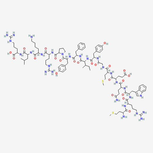
| IUPAC Name | (4S)-4-[[(2S)-5-amino-2-[[(2S)-2-[[(2S)-2-[[(2S)-2-amino-4-methylsulfanylbutanoyl]amino]-5-carbamimidamidopentanoyl]amino]-3-(1H-indol-3-yl)propanoyl]amino]-5-oxopentanoyl]amino]-5-[[(2S)-1-[[2-[[(2S)-1-[[(2S,3S)-1-[[(2S)-1-[[(2S)-1-[(2S)-2-[[(2S)-1-[[(2S)-6-amino-1-[[(2S)-1-[[(1S)-4-carbamimidamido-1-carboxybutyl]amino]-4-methyl-1-oxopentan-2-yl]amino]-1-oxohexan-2-yl]amino]-5-carbamimidamido-1-oxopentan-2-yl]carbamoyl]pyrrolidin-1-yl]-3-(4-hydroxyphenyl)-1-oxopropan-2-yl]amino]-1-oxo-3-phenylpropan-2-yl]amino]-3-methyl-1-oxopentan-2-yl]amino]-3-(4-hydroxyphenyl)-1-oxopropan-2-yl]amino]-2-oxoethyl]amino]-4-methylsulfanyl-1-oxobutan-2-yl]amino]-5-oxopentanoic acid |
| Molecular Formula | C101H152N28O22S2 |
| Molecular Weight | 2174.6 |
| Purity | ≥99% Pure (LC-MS) |
| Liquid Availability | N/A |
| Powder Availability | N/A |
| Storage Condition | Store cold, keep refrigerated. Do NOT freeze. |
| Terms | All products are for laboratory developmental research USE ONLY. Products are not for human consumption. |
**Información Importante: Cada péptido se entrega liofilizado y debe ser reconstituido con Agua Bacteriostática para poder ser administrado en forma líquida.
Vea aquí el video sobre la reconstitución de péptidos
What is MOTS-c?
MOTS-c is a small bioactive peptide derived from mitochondrial DNA that has gathered attention for its unique role in cellular metabolism and stress adaptation. Composed of 16 amino acids, MOTS-c functions as a mitochondrial-derived signaling molecule, influencing nuclear gene expression and coordinating metabolic homeostasis. It is known to activate pathways such as AMP-activated protein kinase, thereby enhancing glucose utilization, promoting fatty acid oxidation, and increasing cellular resistance to oxidative stress. Beyond its metabolic effects, MOTS-c has been studied for its protective roles in age-related disorders, cardiovascular disease, and inflammatory conditions due to its ability to modulate antioxidant responses and reduce cellular apoptosis. Because it is endogenously produced and acts as a regulator of systemic energy balance, MOTS-c has become an important focus of research in understanding how mitochondrial signaling contributes to overall health, disease prevention, and potential therapeutic interventions.
Ver más…
3 Main Research Findings
1) Treatment with MOTS-c was found to reduce high-fat diet-induced and age-related insulin resistance, as well as, diet-induced obesity indicating the potential of the compound to actively regulate metabolic homeostasis.
2) Administration of MOTS-c was shown to prevent the development of heart failure under pressure overload conditions in mice, highlighting the potential therapeutic qualities of the peptide in the treatment of heart failure.
Selected Data
1) Researchers Lee et al used a variety of cell types and animal models to explore both the mechanistic and systemic effects of MOTS-c, while also examining its potential therapeutic applications. Several cell lines were routinely maintained under standard conditions for these studies. HEK293 and HeLa cells were cultured in DMEM, while L6 rat myoblasts were grown in MEM, both supplemented with 10% fetal bovine serum at 37°C in a 5% CO₂ environment. A special subset of HeLa cells lacking mitochondrial DNA, referred to as ρ0 cells, was generated through prolonged exposure to low doses of ethidium bromide. To specifically examine the role of MOTS-c, the team engineered stable cell lines overexpressing MOTS-c: HEK293 cells (MOTS-c-ST) and L6 myoblasts (L6-MOTS-c-ST). These were created by transfecting the cells with a MOTS-c expression construct and selecting with G418 antibiotic. Control cell lines were prepared using an empty vector under the same conditions. Differentiation of L6 myoblasts into mature myotubes was achieved by switching them to a low-serum MEM medium after reaching 80–90% confluence, and maintaining this condition for 8–10 days, with media changes every 2–3 days [1].
Animal studies further explored the systemic impact of MOTS-c administration. All procedures were approved by institutional animal care committees. MOTS-c was synthesized and administered daily to mice through intraperitoneal injections. Both CD-1 (ICR) and C57BL/6 mice were used, obtained from commercial vendors. The animals were placed on either a high-fat diet containing 60% calories from fat, or a matched control diet for eight weeks, with careful monitoring of body weight and food intake. These experiments aimed to model age-related metabolic challenges and assess whether MOTS-c could improve systemic metabolism under such conditions.
Functional assays of mitochondrial activity complemented the transcriptomic data. Real-time measurements of oxygen consumption rate (OCR) were carried out using a Seahorse XF24/96 extracellular flux analyzer to assess mitochondrial respiration. Additional interventions with specific metabolic inhibitors allowed the researchers to determine ATP turnover and maximal respiratory capacity. Parallel measurements of glycolysis were obtained through extracellular acidification rates (ECAR), normalized to basal levels. Cells were sequentially stimulated with glucose, oligomycin, and 2-deoxyglucose (2-DG) to distinguish active glycolysis, maximal glycolytic capacity, and glycolytic reserve. These assays provided a comprehensive view of how MOTS-c expression or treatment affects cellular energy metabolism under different conditions [1].
Overall, this integrated experimental design highlights the connection between mitochondrial genetic integrity, peptide signaling, and systemic metabolic health. The findings support a model in which age-related mitochondrial dysfunction leads to reduced production of MDPs like MOTS-c and humanin, exacerbating metabolic decline. Conversely, supplementation with MOTS-c shows promise in improving metabolic function in aged or metabolically challenged organisms. The recognition of MOTS-c and humanin as functional peptides encoded by the mitochondrial genome also raises the possibility that additional small open reading frames (sORFs) within mitochondria encode other bioactive peptides with regulatory roles. This shift suggests that mitochondria are not merely energy-producing organelles but active participants in organismal homeostasis, with implications for aging, metabolic diseases, and potential therapeutic interventions [1].
2) The study conducted by Zhong et al employed a well-established transverse aortic constriction (TAC) procedure to induce heart failure (HF) in mice. During surgery, animals were anesthetized with 2% isoflurane in oxygen and mechanically ventilated at a rate of 80 breaths per minute with a tidal volume of 0.3 ml. The procedure involved constricting the transverse aorta against a 28-gauge needle using a 7–0 silk suture to generate pressure overload on the heart. Four weeks after the TAC operation, cardiac function was assessed via echocardiography to confirm the development of HF. The mice were then divided into two experimental groups: one group received daily 5 mg/kg subcutaneous administration of the human MOTS-c peptide for four weeks using ALZET osmotic minipumps, while the control group received saline. At the end of the treatment period, mice were euthanized using an overdose of sodium pentobarbital, and heart tissues were harvested and immediately snap-frozen in liquid nitrogen for further analysis [2].
Cardiac function was evaluated using echocardiography at various time points following TAC surgery. The procedure was performed with mice under light anesthesia using an echocardiographic system equipped with a 15-MHz transducer. Short-axis M-mode recordings at the level of the papillary muscles were obtained to evaluate left ventricular (LV) function. For each mouse, parameters were recorded for five consecutive cardiac cycles, and average values were calculated. Primary measurements included the left ventricular internal diastolic diameter (LVIDd), which reflects LV chamber size, and left ventricular ejection fraction (LVEF), an important indicator of systolic function.
Histological analyses were conducted to assess structural cardiac changes, including fibrosis and apoptosis. Hearts were fixed in 10% formalin, embedded in paraffin, and sectioned transversely into 5-μm slices near the apex. Fibrotic remodeling was assessed using Sirius Red staining, which identifies collagen deposition. Interstitial fibrosis was quantified as the percentage of Sirius Red-positive area across 10 randomly selected fields, while perivascular fibrosis was calculated as the ratio of Sirius Red-positive area surrounding vessels to total vascular area. Apoptosis was measured using the TUNEL (TdT-mediated dUTP nick end labeling) assay, and TUNEL-positive cells were visualized with a fluorescence microscope. The proportion of apoptotic cells was determined by dividing the number of TUNEL-positive cells by the total number of cells in four random fields. All histological images were quantified using Image-Pro Plus 6.0 software [2].
Gene expression analyses were carried out using quantitative real-time PCR (qPCR). Total RNA was extracted from heart and other relevant tissues using TRIzol reagent, followed by reverse transcription to complementary DNA (cDNA). The study focused on several key genes, including those encoding fibrotic markers. Gene expression was normalized to β-actin as the internal control, and relative fold changes were calculated [2].
For protein-level analyses, Western blotting was used to assess signaling pathways and protein expression changes associated with MOTS-c treatment. Total protein was extracted from ventricular tissues and cultured H9C2 cardiomyoblast cells. Protein concentration was determined using a BCA assay. Equal amounts of 50 μg of protein were separated by SDS-PAGE, transferred to PVDF membranes, and probed with primary antibodies overnight at 4°C. The antibodies targeted phosphorylated and total AMPK (pAMPK and AMPK), HO-1, and tubulin. After incubation with HRP-conjugated secondary antibodies, protein bands were visualized using ECL reagents. Densitometric analysis was conducted using ImageJ software, with results normalized to the appropriate controls.
The study also included in vitro experiments using H9C2 cells, an embryonic rat heart-derived cell line. Cells were maintained in DMEM/F12 medium supplemented with 5% fetal bovine serum, 100 U/mL penicillin, and 100 mg/mL streptomycin under standard culture conditions. For transfection studies, cells were transfected with either an empty control vector or a MOTS-c expression vector using Lipofectamine 3000 according to the manufacturer’s instructions. Following transfection, cells were cultured for an additional three days in medium with reduced serum.To model oxidative stress, hydrogen peroxide was added at varying concentrations for 24 hours. Cell viability was then assessed using the CCK-8 assay, which measures metabolic activity via absorbance at 450 nm. Cell survival was expressed relative to untreated control cells transfected with the empty vector [2].
Overall, these methods describe a comprehensive experimental design combining in vivo and in vitro approaches to evaluate the therapeutic potential and mechanistic effects of the MOTS-c peptide on cardiac dysfunction, fibrosis, inflammation, and oxidative stress in a pressure overload-induced HF model. The integration of echocardiography, histology, molecular biology, and cell culture techniques provided a multifaceted assessment of MOTS-c’s effects at functional, structural, and molecular levels [2].
Discussion
1) MOTS-c translation occurs in the cytoplasm using the standard genetic code, since mitochondrial translation would produce premature stop codons. This implies that its transcript is exported from mitochondria, a process not fully understood. Sequence alignments across 14 species revealed that MOTS-c is highly conserved, particularly its first 11 amino acids, with some residues showing evidence of positive selection and others under purifying selection. This evolutionary conservation suggested functional significance [1].
To confirm that MOTS-c originates from mitochondrial rather than nuclear DNA, researchers Lee et al performed multiple analyses. BLAST searches revealed no nuclear DNA homologs that match mitochondrial MOTS-c completely, and expression sequence tag databases indicated all transcripts matched the mitochondrial 12S rRNA locus. Rats lack NUMTs for MOTS-c entirely, making the mitochondria its exclusive source. In humans, depletion of mitochondrial DNA in HeLa cells (ρ0 cells) abolished 12S rRNA and MOTS-c expression, while nuclear genes remained unaffected. Similarly, selective depletion of mitochondrial RNA using actinonin eliminated MOTS-c transcripts. Together, these findings demonstrate MOTS-c’s mitochondrial origin. MOTS-c protein was detected in various tissues in mice and rats and in circulation in both species and humans. Interestingly, fasting reduced MOTS-c levels in metabolically active tissues and plasma, but not in homeostatic tissues like the brain and heart [1].
Microarray analyses showed that MOTS-c is bioactive and regulates gene expression. In HEK293 cells treated with MOTS-c, gene expression changed significantly within 4 hours and more distinctly by 72 hours, particularly affecting metabolic and inflammatory pathways. Global metabolomics confirmed these effects, showing large-scale changes in numerous metabolites over time. Many altered metabolites were related to purine, dipeptide, acylcarnitine metabolism, and the methionine cycle. MOTS-c appeared to target the folate–methionine cycle and the connected de novo purine synthesis pathway. It decreased levels of 5-methyltetrahydrofolate and methionine, increased homocysteine, and blocked purine biosynthesis, leading to substantial accumulation of AICAR, a known AMPK activator. MOTS-c treatment triggered phosphorylation of AMPK and related metabolic regulators in a time- and dose-dependent manner, mimicking some effects of drugs like metformin. These findings linked MOTS-c to AMPK activation despite relatively high cellular energy levels.
Metabolic flux analyses revealed that MOTS-c promotes glucose utilization. MOTS-c expressing or treated cells showed increased glucose clearance, lactate production, and enhanced glycolytic flux. Evidence suggested glucose routing toward the pentose phosphate pathway, providing precursors for purine synthesis. Real-time glycolysis measurements confirmed that MOTS-c boosts glycolytic capacity, an effect reversible with folic acid supplementation, consistent with its impact on the folate cycle. Mutated or scrambled MOTS-c peptides lacked these effects, confirming sequence specificity. Knockdowns of AMPKα and SIRT1, as well as pharmacological inhibitors, partially attenuated MOTS-c’s glycolytic effects, implicating these signaling nodes in its mechanism [1].
MOTS-c also suppressed mitochondrial respiration, consistent with the Crabtree effect, where high glycolysis suppresses respiration. This suppression was reversible with folic acid and absent in scrambled or mutant peptides. MOTS-c reduced oxidative capacity and altered TCA cycle metabolites, suggesting that respiratory suppression is secondary to increased glucose uptake. In terms of lipid metabolism, MOTS-c expressing cells displayed higher carnitine shuttle components, reduced essential fatty acids, and increased β-oxidation intermediates, indicating enhanced fatty acid utilization. These effects occurred even under reduced respiration, similar to known AMPK activators [1].
To explore systemic effects, the researchers administered MOTS-c to mice. Acute treatment modestly reduced body weight, food intake, and blood glucose. Circulating inflammatory markers, such as IL-6, TNFα, also decreased. More importantly, MOTS-c improved glucose tolerance and insulin sensitivity, as demonstrated by glucose tolerance tests and hyperinsulinemic-euglycemic clamp studies. MOTS-c enhanced insulin-stimulated glucose disposal primarily in skeletal muscle, where it increased Akt activation and GLUT4 expression. Skeletal muscle and plasma MOTS-c levels naturally decline with age and insulin resistance; treatment of older mice restored insulin sensitivity to youthful levels. In vitro, L6 myocytes overexpressing MOTS-c mirrored these glycolytic enhancements.
Finally, MOTS-c prevented obesity and insulin resistance in mice fed a high-fat diet . While it did not affect body weight under normal diets, MOTS-c dramatically blocked weight gain and hyperinsulinemia in high fat diet-fed mice without altering caloric intake. Hepatic lipid accumulation decreased, and skeletal muscle showed increased AMPK activation and GLUT4 expression. MOTS-c increased glucose utilization and energy expenditure, as evidenced by a higher respiratory exchange ratio and heat production. These findings suggest that MOTS-c improves insulin sensitivity, enhances carbohydrate utilization, and increases energy expenditure, thereby preventing diet-induced obesity [1].
2) The study investigates the therapeutic potential of the mitochondrial-derived peptide MOTS-c in protecting against HF and cardiac injury caused by chronic pressure overload. HF induced by pressure overload is associated with progressive cardiac dysfunction, structural remodeling, fibrosis, inflammation, oxidative stress, and cardiomyocyte apoptosis. To explore whether MOTS-c could mitigate these detrimental effects, researchers used a well-established mouse model of heart failure induced by TAC, as well as in vitro experiments in cultured cardiac cells [2].
In the first part of the study, the researchers examined the impact of MOTS-c administration on cardiac function under pressure overload. TAC surgery was performed to induce heart failure, and MOTS-c treatment was initiated via osmotic pump four weeks post-surgery. Cardiac function was monitored over time by echocardiography, assessing key parameters including LVEF and LVIDd. The TAC procedure successfully induced cardiac dysfunction and ventricular dilation by week four, confirming the validity of the model. Without treatment, heart function continued to deteriorate significantly by week eight. However, mice treated with MOTS-c exhibited a marked attenuation of cardiac dysfunction and structural deterioration compared to untreated TAC mice. Importantly, heart rate and body temperature remained unchanged across groups, indicating that the benefits observed were specific to cardiac function rather than systemic physiological changes. These findings suggest that MOTS-c has therapeutic potential for pressure overload-induced heart failure [2].
Subsequent analyses explored the effects of MOTS-c on structural remodeling of the heart, particularly fibrosis and apoptosis, which are hallmarks of heart failure progression. Chronic pressure overload leads to excessive deposition of collagen in the interstitial and perivascular regions of the left ventricle. Sirius Red staining revealed that TAC mice developed extensive myocardial fibrosis compared to control mice. In contrast, MOTS-c treatment significantly reduced fibrosis levels. Consistent with histological findings, mRNA expression of fibrosis-related genes, including collagen I, collagen III, and connective tissue growth factor (CTGF), was elevated in TAC hearts and markedly suppressed by MOTS-c treatment. Similarly, the researchers assessed cardiomyocyte apoptosis using TUNEL staining. TAC mice displayed a significant increase in apoptotic cells, while MOTS-c administration significantly reduced cell death. These results demonstrate that MOTS-c mitigates maladaptive structural remodeling in the overloaded heart by reducing both fibrosis and apoptosis.
Inflammation plays a critical role in the initiation and progression of heart failure, and the researchers next investigated whether MOTS-c exerts anti-inflammatory effects in the heart under pressure overload conditions. mRNA expression levels of key pro-inflammatory cytokines, including TNF-α, IL-1β, and IL-6, were significantly upregulated in TAC mice compared to controls. Treatment with MOTS-c markedly suppressed the expression of these inflammatory genes. Immunohistochemical analysis further confirmed an increased presence of TNF-α in TAC hearts, which was notably reduced by MOTS-c administration. These findings suggest that MOTS-c can attenuate the inflammatory response associated with pressure overload-induced cardiac injury [2].
Oxidative stress is another major contributor to heart failure pathogenesis, often linked to insufficient activation of endogenous antioxidant defense mechanisms. The transcription factor Nrf2 plays a central role in regulating antioxidant gene expression. Previous studies suggested that MOTS-c may enhance antioxidant responses, so researchers Zhong et al examined Nrf2-regulated antioxidant genes in the heart. Interestingly, TAC alone did not alter the expression of key antioxidant genes such as heme oxygenase-1, NAD(P)H quinone dehydrogenase 1, and glutamate-cysteine ligase catalytic subunit. However, MOTS-c treatment significantly upregulated the expression of all three genes. Protein levels of HO-1 were also increased with MOTS-c therapy. Since AMP-activated protein kinase activation is known to mediate MOTS-c’s antioxidant effects, the researchers assessed AMPK activity by measuring phosphorylated AMPK levels. MOTS-c-treated hearts exhibited significantly elevated p-AMPK/AMPK ratios, indicating robust activation of this pathway. These results suggest that MOTS-c enhances antioxidant defense mechanisms in the failing heart through Nrf2-related gene upregulation and AMPK activation.
Finally, to further investigate the cytoprotective effects of MOTS-c and its mechanisms at the cellular level, the researchers conducted in vitro experiments using cultured cardiac H9C2 cells subjected to oxidative stress. A MOTS-c-expressing plasmid was constructed and transfected into H9C2 cells, which also expressed EGFP for visualization of transfection efficiency. Fluorescent microscopy confirmed robust MOTS-c expression. As observed in vivo, MOTS-c overexpression in cardiac cells significantly increased phosphorylated AMPK levels, indicating activation of this protective signaling pathway. To model oxidative injury, the transfected cells were treated with varying doses of hydrogen peroxide for 24 hours, and cell survival was measured using a CCK-8 assay. Cells overexpressing MOTS-c exhibited significantly greater resistance to H₂O₂-induced cell death compared to control cells transfected with an empty vector. These findings reinforce the conclusion that MOTS-c confers protection against oxidative stress-induced apoptosis in cardiac cells, mediated at least in part by AMPK activation [2].
In summary, this study demonstrates that MOTS-c has a multifaceted protective effect on the heart under conditions of chronic pressure overload. Systemically administered MOTS-c improves cardiac function and prevents adverse structural remodeling by reducing fibrosis, apoptosis, and inflammation. Additionally, MOTS-c enhances the heart’s antioxidant capacity by upregulating Nrf2-regulated antioxidant genes and activating AMPK signaling. The in vitro data corroborates these findings by showing that MOTS-c overexpression protects cardiac cells against oxidative injury. Collectively, these results highlight the therapeutic potential of MOTS-c as a novel strategy for treating pressure overload-induced heart failure, warranting further investigation in preclinical and clinical settings [2].
Disclaimer
**LAB USE ONLY**
*This information is for educational purposes only and does not constitute medical advice. THE PRODUCTS DESCRIBED HEREIN ARE FOR RESEARCH USE ONLY. All clinical research must be conducted with oversight from the appropriate Institutional Review Board (IRB). All preclinical research must be conducted with oversight from the appropriate Institutional Animal Care and Use Committee (IACUC) following the guidelines of the Animal Welfare Act (AWA).
Citations
[1] Lee C, Zeng J, Drew BG, et al. The mitochondrial-derived peptide MOTS-c promotes metabolic homeostasis and reduces obesity and insulin resistance. Cell Metab. 2015;21(3):443-454. doi:10.1016/j.cmet.2015.02.009
[2] Zhong P, Peng J, Hu Y, Zhang J, Shen C. Mitochondrial derived peptide MOTS-c prevents the development of heart failure under pressure overload conditions in mice. J Cell Mol Med. 2022;26(21):5369-5378. doi:10.1111/jcmm.17551
MOTS-c is a mitochondrial-derived peptide known for its ability to combat symptoms of physical decline and aging as well as maintain homeostasis in muscle tissues. Mitochondria are directly involved in the age-induced decline of metabolic and physical health as they produce the bulk of energy in the body and by coordinating communication to the nucleus of the cell and various other organelles.
Mitochondria possess specific genomes that are encoded with 13 distinct protein-coding genes as well as various, recently discovered short open reading frames (sORFs). These sORFs are known to produce mitochondrial-driven peptides (MDPs), that adapt and aid in multiple different physiological functions. MOTS-c is an MDP that aids in improving metabolic homeostasis through AMPK and the regulation of nuclear gene expression following translocation. Expression of MOTS-c is highly age-dependent and the peptide is commonly referred to as a “mitochondrial hormone” due to its presence in skeletal muscle and circulating blood (https://www.nature.com/articles/s41467-020-20790-0.)
 
MOTS-c and Physical Performance
Given the previously stated information, researchers Reynolds et. Al conducted a study observing physical performance enhanced by MOTS-c in young (2 months), middle-aged (12 months), and old (22 months) mice. In addition to observing the physical performance of the mice, the researchers were looking for data on metabolic and proteostatic-related nuclear genes, the metabolism of skeletal muscles, and myoblast adaptation to stress.
Reynolds et. Al began the study by administering 5 mg/kg of body weight per day of MOTS-c to 12-week-old mice for 2 weeks. Rotarod tests confirmed that after a week, administration of MOTS-c was able to improve physical performance. This claim was further solidified by a treadmill running test. Since MOTS-c is also capable of improving metabolic homeostasis, the researchers took their study a step further and tested the running performance of young mice when placed under the dietary stress of a high-fat diet (60% of calories from fat).
These mice were given two different doses of MOTS-c, 5 or 15mg/kg/day. It was found that mice receiving higher doses of the peptide showed a significant improvement in both power output and running capacity after 10 days and improved skeletal muscle insulin sensitivity after 7 days. The treadmill test was increased to a sprint to test endurance and 100% of the mice receiving 15 mg/kg/day were able to pass the minimum rate of 23 m/min. Furthermore, NMR analyzers found that in young mice MOTS-c slowed down the fat gain and increased lean mass.
Both middle-aged and old mice were treated with 15 mg/kg/day for 2 weeks and then were made to complete a treadmill running test. In middle-aged and older mice it was found that there was a significant improvement in power output as well as distance for both groups. Additionally, older mice receiving doses of the peptide were submitted to the same sprint (23 m/min) test as the young mice and 17% of them passed the test, while 0% of the control group passed.
In addition to the testing of young, middle-aged, and old mice, the researcher also tested the efficiency of MOTS-c on mice in their end-of-life stages (24 months) to see the peptides’ effects on quality of life. It was found that 15mg/kg 3 times a week improved grip strength, 60-second walk times, and stride length. Overall Reynolds et. Al concluded that MOTS-c has the capability to significantly improve physical performance and capacity in all life stages (https://www.nature.com/articles/s41467-020-20790-0).
PEPTIDES PREFER THE COLD
Keep peptide vials refrigerated at all times to reduce peptide bond breakdown. DO NOT FREEZE. Most peptides, especially shorter ones, can be preserved for weeks if careful.
Always swab the top of the vial with an alcohol wipe or rubbing alcohol before accessing.
ONLY MIX WITH STERILE BACTERIOSTATIC WATER
The purity and sterility of bacteriostatic water are essential to prevent contamination and to preserve the shelf-life of dissolved peptides.
Push the pin through the rubber stopper at a slight angle, so that you inject the bacteriostatic water toward the inside wall of the vial, not directly onto the powder.
Lyophilized peptide should be stored at -20°C (freezer), and the reconstituted peptide solution at 4°C (refrigerated). Do not freeze once reconstituted.
NEVER SHAKE A VIAL TO MIX.
Air bubbles are unfavorable to the stability of proteins.
MOTS-c is sold for laboratory research use only. Terms of sale apply. Not for human consumption, nor medical, veterinary, or household uses. Please familiarize yourself with our Terms & Conditions prior to ordering.


Valoraciones
No hay valoraciones aún.